| Step 7: What tests might be ordered? |
Your doctor can use a number of helpful imaging studies to examine the structures of your back, including your spine, if needed. Other tests assess the electrical activity of your muscles and nerves. These tests are used to try to identify the exact location and source of your pain.
X-Ray
An x-ray of your back may show a sign of an injury, infection, fracture, osteopenia (loss of bone density), or tumor. However, most people with low back pain due to strain or spinal problems have normal x-rays. If the results of an x-ray are not definitive, your doctor may order a CT or MRI scan.
CT and MRI scans
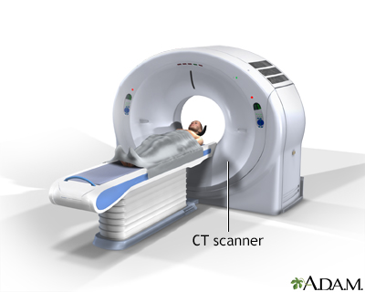
Computed tomography (CT) and magnetic resonance imaging (MRI) can be used to identify disk abnormalities and any other problems in the back. MRIs are more accurate for soft tissue, but CTs are better for bones and small joints. MRIs provide very clear pictures of all parts of the back, including muscles, ligaments, and the vertebrae. These tests can also identify infections and tumors if present.
Your doctor may order a lumbar MRI if you have signs or symptoms of:
- Low back or leg pain that is severe or does not get better after treatment by your doctor
- Low back pain and weakness, numbness, or other unusual findings on physical exam
- Fever or other signs of infection
- Injury or trauma to the lower spine
- Problems with bowel or bladder function

CT scanMRI
Nerve and muscle studies
Based on your description of back pain, your physical exam, or any imaging studies performed, your doctor may order studies to test the activity of your back muscles and spinal nerves. Nerve conduction studies are done more often than muscle testing. (Muscle testing can be quite painful.) Nerve conduction tests involve placing electrodes on your skin and applying small electrical signals. With these shocks, the speed with which your nerves conduct the signal is measured.
Other tests
Blood and urine samples may be used to test for infection, arthritis, or other conditions.
Reviewed By: Andrew W. Piasecki, MD, Camden Bone and Joint, LLC, Orthopaedic Surgery/Sports Medicine, Camden, SC. Review provided by VeriMed Healthcare Network. Also reviewed by David Zieve, MD, MHA, Medical Director, A.D.A.M., Inc.
© 1997- A.D.A.M., a business unit of Ebix, Inc. Any duplication or distribution of the information contained herein is strictly prohibited.
