| Step 4: How osteoarthritis is diagnosed |
Older people often don't realize that they have osteoarthritis if they are free of pain and other symptoms. However, x-rays often reveal some osteoarthritis of the spine or fingers in elderly individuals.

Your doctor can diagnose osteoarthritis with:
- A physical examination and discussion of your symptoms
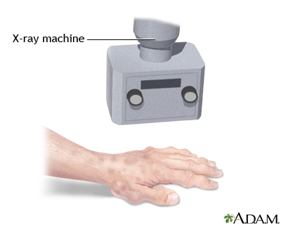
- X-rays and possibly other imaging studies
- Fluid aspiration and blood analysis
Physical examination
If you have joint pain or other joint symptoms, you can talk to your primary care provider, who will be very familiar with diagnosing arthritis. You can also see a rheumatologist (a specialist in joint disorders) or an orthopedic surgeon. The doctor will ask you about any illnesses, injuries, or operations you have had in the past. You should tell the doctor about any allergies or other conditions you have right now.
The doctor will inspect your joints, checking for swelling, deformity, redness, heat, tenderness, and rashes. The doctor will want to identify which joints are affected, and how, in order to distinguish osteoarthritis from other forms of arthritis.
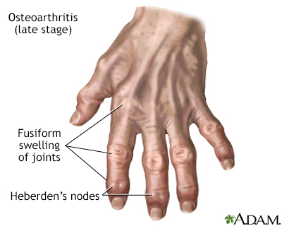
The muscles that surround painful, underused joints may be weak. The symptoms in your hands may be especially important in making the diagnosis. Osteoarthritis tends to involve the base of the thumb and the middle and end joints of the fingers.

| Osteoarthritis is associated with the aging process and can affect any joint. The cartilage of the affected joint is gradually worn down, eventually causing bone to rub against bone. Bony spurs develop on the unprotected bones causing pain and inflammation. |
X-rays and imaging studies
X-rays can confirm that you have arthritis, but will not necessarily indicate the type of arthritis. Your doctor will look for specific structural changes in the joints that suggest osteoarthritis, such as:
- Narrowing of the joint space. This occurs due to loss of cartilage (for example, joint space narrowing of the inside half of the knee).
- Bony spurs. These are outgrowths of new bone that develop at the margin of the joint. It is nature's way of protecting the joint.
- One-sided distribution (for example, one knee, one hip) of joint irregularities.
- Cysts. These may be seen in the bone just beneath the joint surfaces.
By contrast, imaging studies in people with rheumatoid arthritis more often shows:
- Loss of calcium from the bone (localized bony decalcification)
- Erosion-producing defects or holes in the bones in a joint
- Changes in many joints on both sides of the body
Laboratory tests
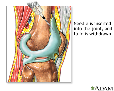
If there is a question about the exact nature of joint swelling, the physician may perform a joint aspiration. During this procedure:

- A needle is gently inserted into the joint to withdraw a small amount of synovial fluid from the joint.
- The fluid then is tested for chemistry, viscosity (thickness), blood cell counts, overall appearance, and microorganisms (if an infection is suspected).
- The fluid from an osteoarthritis joint is usually clear, whereas in rheumatoid arthritis, it is cloudy due to the presence of many white blood cells.
- The fluid then is tested for crystals to exclude diagnoses such as gout.
- Sometimes the fluid from an osteoarthritis joint contains crystals such as calcium pyrophosphate, which may cause mild irritation and increase swelling.
Blood tests may be ordered to identify infection, measure blood cell counts, and pinpoint telltale diagnostic findings such as rheumatoid factor (RF), which are more common in people with inflammatory types of arthritis, such as rheumatoid arthritis.
Blood and urine tests may be ordered to rule out conditions such as gout. The blood from people with gout contains a high level of uric acid, which is associated with the buildup of arthritis-causing crystals in the joint fluid.
Did you know...?
|
Reviewed By: Ariel D. Teitel, MD, MBA, Clinical Associate Professor of Medicine, Division of Rheumatology, NYU Langone Medical Center. Review provided by VeriMed Healthcare Network.
